
Unusual things, found and filed.Independent web · Since 2026
Odd Almanac
Search

Small wonders in the natural world

Ice, living processes and the experiments that help explain their forms.

15 entries · 4 sections

Editorial illustration: a branching tree of paths
Editorial illustration · Z13 visual study

A small directory of unusual natural phenomena. Observations, experimental findings and proposed explanations retain their own context.

Ice on dead wood

A study of fine ice filaments distinguishes a freezing mechanism from fungal shaping.

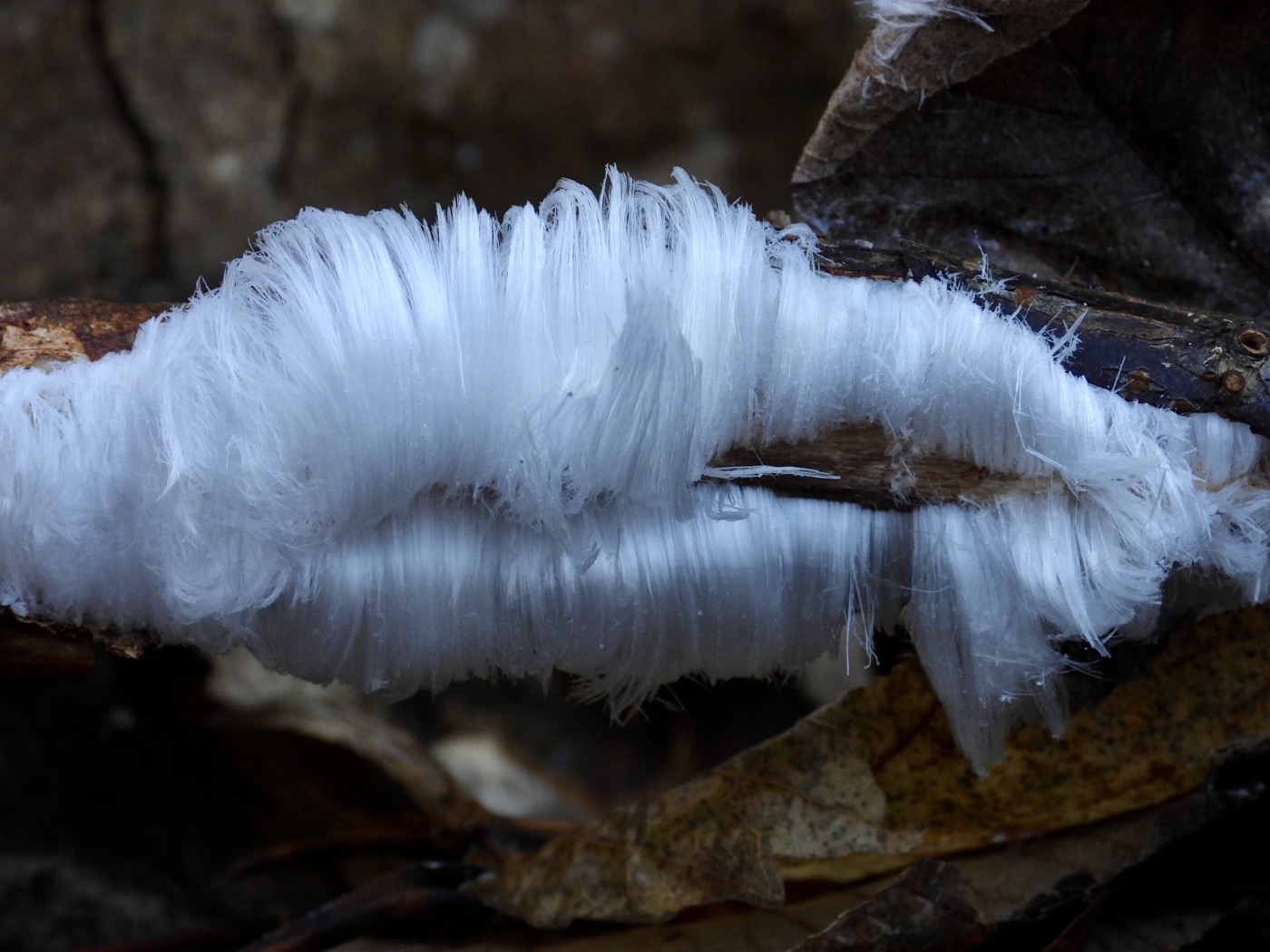
Fine white ice filaments curl from a dark fallen branch above brown leaves.
Hair ice on dead wood in Harju County, Estonia, photographed by Hall Wild on 22 November 2025. This later observation is separate from the wood examined in the 2015 study. Hall Wild / Wikimedia Commons · CC BY 4.0

Research & assessmentOriginal paper · 22 July 2015

The fungus that helps ice grow into hair

On damp dead wood, fine ice filaments depend on fungal activity; the exact stabilising substance remains an open question.

Added · Daily discovery

Fine white curls can appear on dead branches during damp weather just below freezing. They are ice, growing out from the wood. The phenomenon is called hair ice; a 2015 investigation separated the process that freezes the water from the biological activity that gives the ice its shape.

Diana Hofmann, Gisela Preuss and Christian Mätzler reported their work in Biogeosciences on 22 July 2015. Their experiments supported ice segregation: water within the wood supplies ice forming at its surface. Samples with and without active fungus showed similar temperature changes during freezing. Fungal heat was too small to explain the process.

The difference was the shape. Without fungal activity, the ice formed a crust rather than fine hairs. The researchers found Exidiopsis effusa on every hair-ice-producing wood sample they examined. Their evidence linked this winter-active fungus to the delicate structure, rather than establishing a new way for water to freeze.

The chemistry remained less settled. Melted hair ice contained organic compounds associated with decomposed wood. The authors proposed that a substance linked to the fungus prevents the filaments from merging into larger crystals. Their paper left the precise recrystallisation inhibitor to be identified.

The European Geosciences Union’s accompanying account traces the fungal suggestion back to Alfred Wegener in 1918. It also explains why the ice is easy to miss: it often grows overnight and disappears as the morning warms.

Hall Wild’s photograph records hair ice in Harju County, Estonia, on 22 November 2025. It illustrates the phenomenon; it is a later observation, separate from the wood examined in the 2015 study.

A 2015 experimental study and its institutional account. The stabilising chemical was unresolved; the later photograph is not a sample from those experiments.

Source details & what was checked
  • Diana Hofmann, Gisela Preuss and Christian Mätzler · 22 July 2015
    Publisher abstract, citation and publication dates read on desktop and phones. The abstract reports fungal suppression tests, Exidiopsis effusa on investigated samples, temperature comparisons, ice segregation and an unidentified recrystallisation inhibitor. The offered Article PDF was successfully downloaded through the normal publisher link on a phone-width browser. This entry does not claim a full paper read or experimental reproduction.
    Evidence for biological shaping of hair ice (external source, opens a new tab) · In the source library
  • European Geosciences Union · 22 July 2015
    Complete institutional release read, including the named researchers, earlier Wegener suggestion, microscopic sample findings, distinction between physical freezing and fungal shaping, tentative inhibitor explanation and fleeting winter conditions. This is the journal publisher’s account of the same study, not an independent replication. Linked videos and other branches were not inspected.
    Fungus shapes hair ice (external source, opens a new tab) · In the source library
Back to sections ↑

Shapes, light and living systems

Natural forms with specific physical or biological explanations, and the observations behind them.

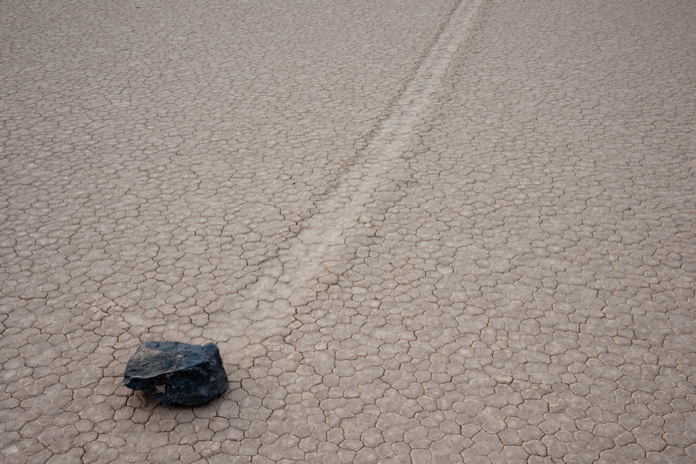
A stone with a long track across the cracked surface of Racetrack Playa.
A stone and its trail at Racetrack Playa, Death Valley, 21 February 2011. Photographed before the 2013–14 movement observations discussed here. Jason Hickey · CC BY 3.0

History & recordFirst-party institutional account · 27 August 2014; page updated 28 February 2015

The desert stones pushed by thin sheets of ice

At Racetrack Playa, direct observations in 2013 caught floating ice panels pushing stones across wet mud.

A stone can leave a long track across the dry floor of Racetrack Playa in Death Valley. For decades, the tracks were easier to find than a moving stone. An experiment set up in 2011 finally caught the process during the winter of 2013–14.

The National Park Service’s August 2014 account describes a weather station and fifteen GPS-equipped rocks brought in from outside the park. Researchers arrived in December 2013 to find a shallow pond, overnight ice and, eventually, moving stones.

Sunlight broke the thin ice into floating panels. Light winds moved those panels, which pushed exposed rocks over soft mud. The observed motion was slow enough to be hard to notice without a fixed reference point.

This was a measured explanation for particular movement events, not a claim that every stone had been tracked. The researchers specifically said they had not yet watched the largest rocks move.

Source details & what was checked
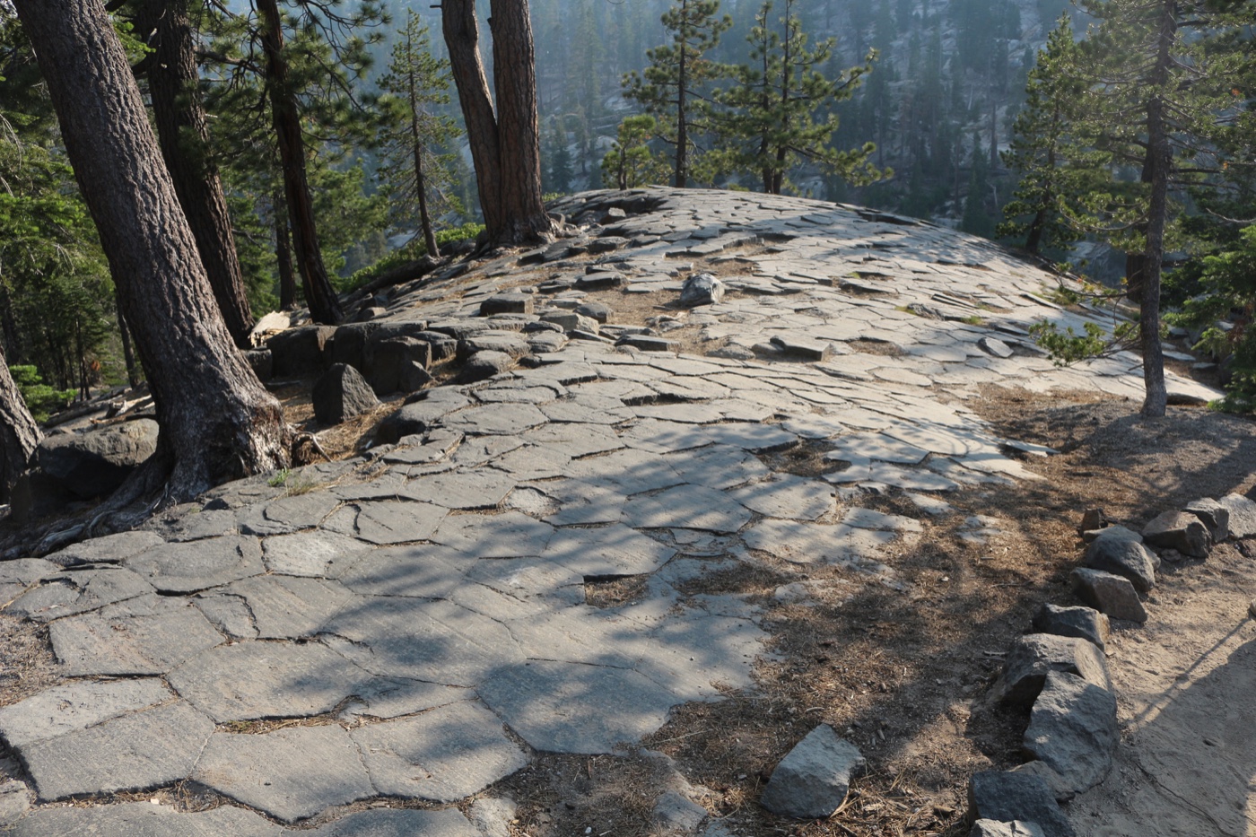
Polygonal tops of basalt columns at Devils Postpile.
The tops of basalt columns at Devils Postpile National Monument, California, 3 July 2018. Lucas·G · CC BY-SA 4.0

History & recordFirst-party institutional account · Updated 18 April 2023

The stone columns made by cooling cracks

Basalt can split into polygonal columns as a hot flow contracts; the cracks follow its cooling surfaces.

Volcanic rock can split into columns that resemble a tightly packed pavement viewed from above. The National Park Service explains these shapes as columnar joints: regularly spaced fractures that divide a rock body into roughly prismatic pieces.

The fracture pattern develops after the rock has solidified but remains hot. Continued cooling causes contraction. Cracks advance inward from cooling surfaces, with columns oriented perpendicular to those surfaces rather than necessarily pointing straight upwards.

Six-sided columns are common because three fractures meeting at roughly 120-degree angles efficiently relieve contractional stress. They are not compulsory: the same account describes columns with three to seven sides, alongside irregularly jointed zones within some flows.

The explanation extends beyond basalt to other volcanic deposits and shallow intrusions. Its park examples include Devils Postpile, where about 55 percent of the columns are hexagonal. Even this celebrated formation contains curved and tilted columns; the source presents their shape as evidence about cooling, not a perfectly repeated geometric mould.

Source details & what was checked
  • National Park Service, Volcanoes, Craters & Lava Flows · Updated 18 April 2023
    Complete Introduction and How Columnar Jointing Forms, park examples and updated date meaningfully read with web reader. No named prose author supplied on exact page; graphics credited Allyson Mathis. No new field experiment or current forecast claimed. Independent destination desktop/mobile QA pending.
    Columnar Jointing (external source, opens a new tab) · In the source library
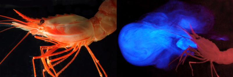
Paired photographs of an orange shrimp and the same shrimp beside a cloud of bright blue light.
The deep-sea shrimp Heterocarpus ensifer and the same animal releasing luminous material near its mouth. These paired photographs accompanied NOAA’s Bioluminescence and Vision 2015 expedition report; they show a different organism from the dinoflagellates in glowing bays. Sönke Johnsen and Katie Thomas / NOAA Ocean Explorer · CC BY-SA 2.0

History & recordFirst-party institutional account · Updated 23 September 2026

When the sea makes its own light

Marine organisms produce light through chemical reactions; glowing bays are one visible example of a much wider phenomenon.

A dark sea can contain its own sources of light. NOAA’s explanation of bioluminescence describes living organisms releasing light through chemical reactions, inside their bodies or through material they eject.

The phenomenon spans marine habitats, from surface waters to the deep seabed. NOAA’s separate account of Mosquito Bay on Vieques identifies the tiny dinoflagellate Pyrodinium bahamense as the organism behind that bay’s night-time display.

Light can have different jobs in different organisms. NOAA lists communication, attracting or detecting prey, and warning or evading predators among the functions known to researchers. It also says that the function is unresolved in some animals.

Bioluminescence differs from fluorescence, in which incoming light is absorbed and emitted at another colour. The two reading routes give a general mechanism and a particular place, without treating every luminous marine image as the same species or the same experiment.

Source details & what was checked
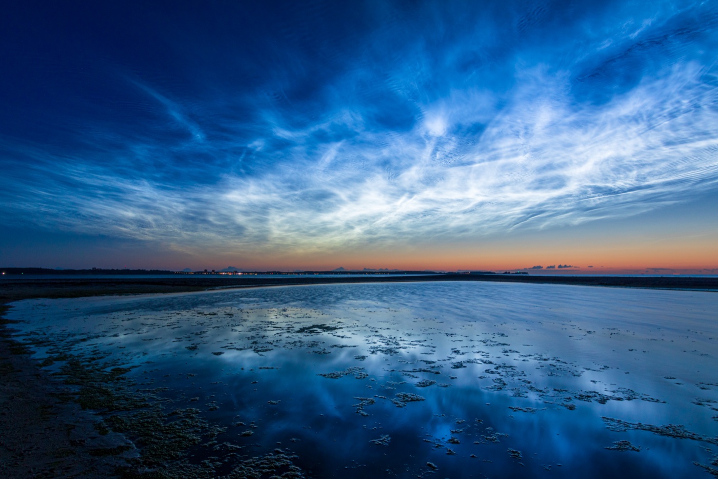
Luminous pale-blue clouds above a twilight horizon and water.
Noctilucent clouds over the Baltic Sea from Laboe, Germany, 21 June 2019. A ground-level photograph, not the AIM satellite map discussed here. Matthias Süßen · CC BY-SA 4.0

History & recordFirst-party institutional account · 6 May 2008

The clouds that catch sunlight after sunset

Noctilucent clouds are high-altitude ice clouds, lit by the Sun while the lower sky is already dark.

The Sun can have set for an observer while still lighting clouds far above the ordinary weather. Noctilucent, or night-shining, clouds sit near the top of the atmosphere’s mesosphere and contain ice particles.

NASA’s 2008 account describes early observations from its AIM satellite. One image, collected on 11 June 2007, mapped the northern polar region: white represented denser ice particles, blue represented lower densities, and black marked missing data. It was a scientific map, not a photograph of a blue-glowing polar landscape.

The mission found clouds that changed over hours and days. Its early measurements placed the ice in a high, comparatively thin atmospheric layer. Their altitude lets them reflect sunlight after lower clouds have entered shadow.

The account records the questions and findings of the early AIM mission. It offers a way to recognise an unusual form of reflected sunlight, while keeping its dated observations separate from claims about present-day cloud frequency.

Source details & what was checked
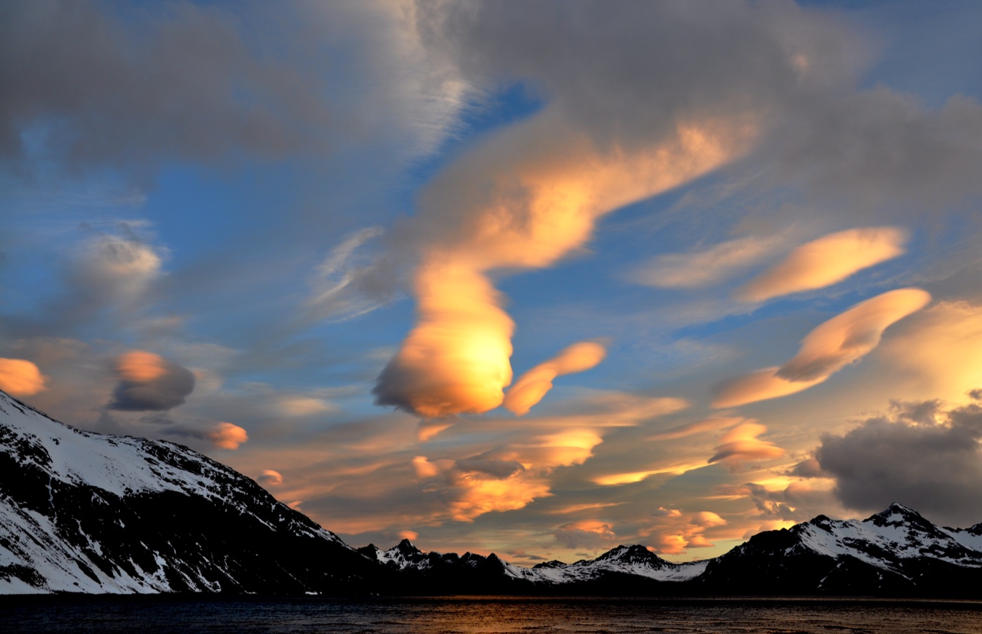
Smooth lens-shaped clouds above the mountains of South Georgia at sunset.
Lenticular clouds at sunset over Leith Harbour, Stromness Bay, South Georgia, 2010. A contextual example; the cited weather-service examples are from New Mexico. Serge Ouachée (Butterfly austral) · CC BY-SA 3.0

History & recordFirst-party institutional account · Undated feature; examples from 2006–2010

The cloud that stays put while air passes through

Lenticular clouds can keep their position because droplets form and disappear within a standing atmospheric wave.

A smooth cloud can seem parked above a mountain even when the air is moving quickly. The National Weather Service’s Albuquerque office explains this apparent contradiction through mountain waves.

When stable air crosses a suitably aligned ridge, it can rise and fall in waves downwind. If there is enough moisture, cloud develops in the rising part of a wave. Near and beyond the crest, the cloud is continually forming and dissipating.

The visible shape can therefore stay close to one place while air travels through it. The office calls these altocumulus standing lenticular clouds and illustrates the explanation with dated New Mexico photographs and a weather sounding.

The feature is also a reminder that a handsome cloud has a physical setting: mountain waves can produce severe turbulence. This is an explanation of the cloud’s form, not a current forecast or flying guide. The page’s historical examples should be read with their own dates.

Source details & what was checked
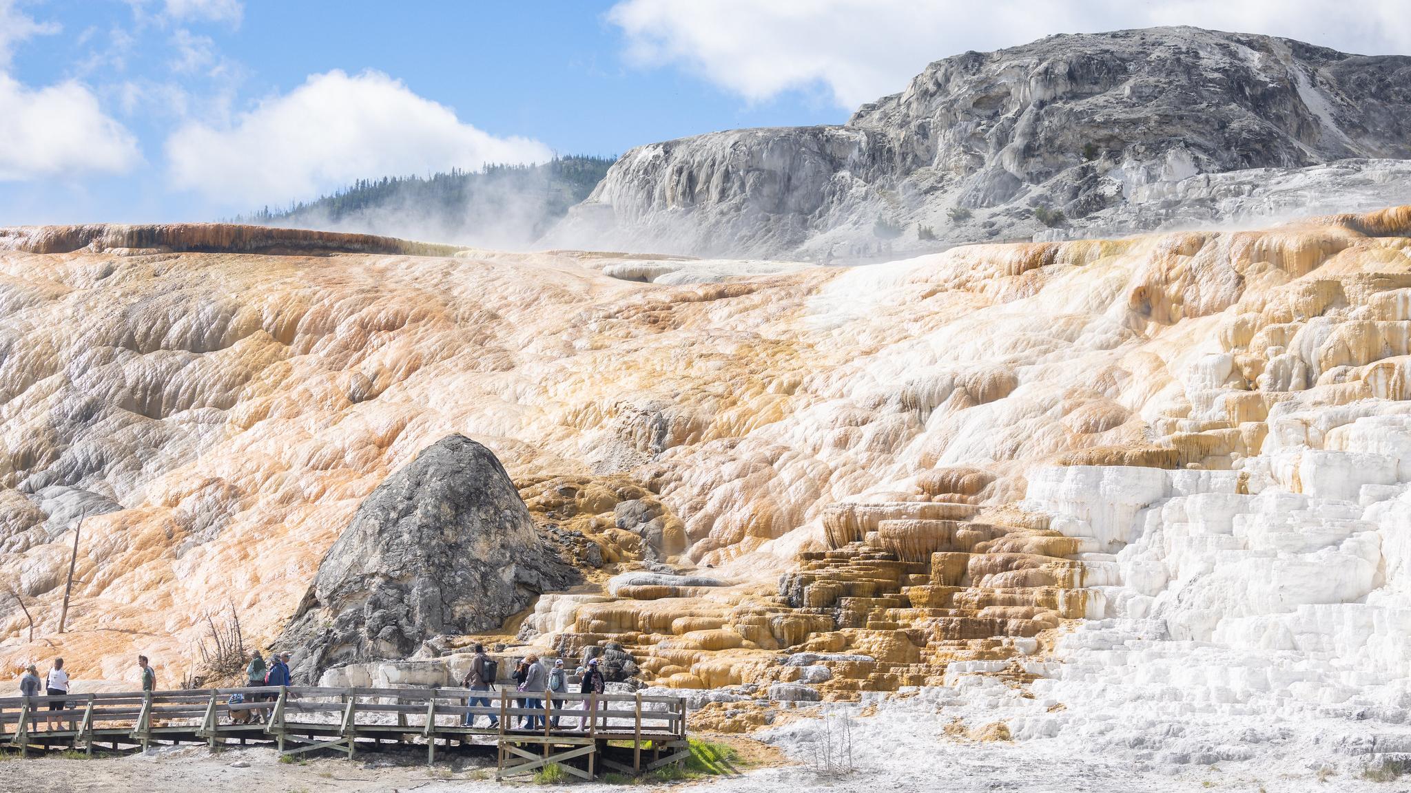
Steaming pale travertine terraces above a boardwalk at Mammoth Hot Springs.
Mammoth Hot Springs travertine terraces, photographed by Jacob W. Frank for the National Park Service. This is the published site view, with its supplied frame preserved. Jacob W. Frank / National Park Service · Public domain — U.S. Government work

History & recordFirst-party institutional account · Updated 27 November 2024

The hot springs that build their own terraces

At Mammoth Hot Springs, dissolved limestone returns as travertine when the rising water releases carbon dioxide.

The pale terraces at Mammoth Hot Springs in Yellowstone are made by water depositing rock at the surface. Their appearance is temporary: as the spring system changes, calcium carbonate continues to accumulate in new forms.

The National Park Service traces the water to rain and snow in surrounding mountains. Underground channels carry it through limestone, where it acquires dissolved calcium carbonate. When the water reaches the surface and releases carbon dioxide, that material can be deposited as travertine.

The colours add a second process. Communities of heat-loving microorganisms occupy the hydrothermal features. The park’s account describes different colours associated with organisms living at different temperatures.

The same source preserves an unresolved detail: the precise volcanic heat source at Mammoth has more than one proposed explanation. The mineral-deposition account is firmer than a single settled diagram of the plumbing beneath it. The linked park record explains the formation without presenting the terraces as permanent sculptures.

Source details & what was checked
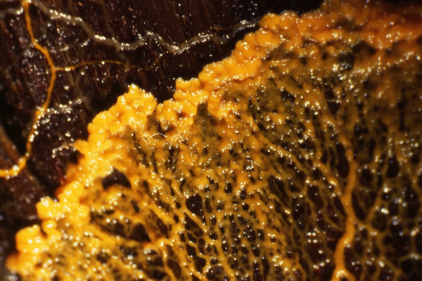
A yellow Physarum polycephalum plasmodium spreading in branching veins.
A Physarum polycephalum plasmodium, photographed 7 April 2013. A contextual image, not a specimen from the 2021 experiment. HelenGinger · CC BY-SA 3.0

Research & assessmentOriginal paper · 22 February 2021

The slime mould that reshapes its tubes around food

A food stimulus reorganises a slime mould’s living tubes; a study separates measured changes from a proposed flow-based mechanism.

Physarum polycephalum is a single-celled organism whose body forms an interconnected network of tubes. When it encounters food, some tubes widen while others shrink, changing how material flows through the network.

In a 2021 study, Mirna Kramar and Karen Alim followed that reorganisation after a local nutrient stimulus. They observed thicker tubes near the stimulus and relative shrinkage elsewhere. A flow model linked those changes to transport of a substance that softens tube walls.

The new diameter pattern emerged within about 15 minutes and persisted through the 45-minute observation period. The study’s discussion says the identity of the proposed softening chemical remained unknown. Its mechanism combines measurements with a theoretical model.

The authors call the network change a way of encoding memory. Their paper also explains the difficulty of comparing two identical networks with different histories. That interpretation should remain attached to the researchers and their experimental arrangement, rather than becoming a general claim about a slime mould’s inner experience.

Measured tube changes and a model-based explanation; the authors’ memory interpretation is attributed and the chemical agent remained unidentified.

Source details & what was checked
Snow among orange hoodoo spires at Bryce Canyon in early light.
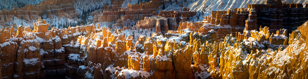
Snow among Bryce Canyon’s hoodoos in Brian Roanhorse’s NPS photograph. The panoramic frame is preserved as published; no capture date is inferred from the filename. Brian Roanhorse / National Park Service · Public domain — U.S. Government work

History & recordFirst-party institutional account · Updated 4 September 2021

The rock spires sculpted by freezing water

Bryce Canyon’s hoodoos emerge as ice and rain progressively dismantle uplifted sedimentary rock.

A hoodoo is a narrow rock spire with an uneven outline. At Bryce Canyon in Utah, crowds of these pillars are remnants of a landscape being taken apart, rather than separate towers built from the ground up.

The National Park Service explains their formation as a sequence: sediments accumulated in an ancient lake and floodplain, the resulting rocks were uplifted, and weathering and erosion began to sculpt them at high elevation.

Water entering cracks can freeze and expand, forcing the rock apart. Over time, broad rock walls develop openings; the openings enlarge and leave isolated pillars. Rain also dissolves the calcium-carbonate cement in the rocks, with differences between layers helping produce their irregular profiles.

The park’s explanation gives both the long geological setting and the smaller repeated processes that alter a single spire. It distinguishes weathering, which breaks rock down, from erosion, which carries the loosened material away. Both processes contribute to the changing landscape.

A dated NPS geological interpretation; current site conditions and individual formation ages are not independently measured here.

Source details & what was checked
  • National Park Service / Bryce Canyon National Park · Updated 4 September 2021
    Complete three-part geological interpretation read: sediment deposition, uplift, frost wedging, wall/window/pillar sequence and calcium-carbonate dissolution. No present-day weather forecast or physical visit claimed.
    Hoodoos (external source, opens a new tab) · In the source library
Round green marimo algae displayed underwater in a museum tank.
Marimo on display at the Akankohan Eco-Museum Center, Japan. The original Japanese description dates the photograph to March 2005; it does not show the 2021 field study. コムケ at Japanese Wikipedia · CC BY-SA 3.0

Research & assessmentOriginal paper · 10 November 2021; repository issue 23 October 2026 is not the publication date

The green lake balls polished by waves

Marimo are algal aggregates; a Lake Akan study connected their spherical form with wave-driven rotation and water exchange.

A marimo looks like a neatly made green ball, but it is an aggregate of the filamentous freshwater alga Aegagropila linnaei. At Lake Akan in Japan, some grow to an unusually large size.

A 2021 study led by Keisuke Nakayama examined the physical environment behind that shape. The researchers linked wind-driven waves to the rotation that polishes the aggregates into spheres. Their account also connects this motion with exchange of water and nutrients between the balls’ interiors and the surrounding lake.

The paper reports alternating internal bands, which the authors interpret as annual rings related to seasonal ice cover. These findings belong to the studied Lake Akan environment; they are not a universal care recipe for every ball sold as a marimo.

The Japan Tourism Agency’s English interpretation adds an accessible explanation of tumbling and uneven wave strength. Read together, the sources show a biological form maintained through interaction with moving water, while preserving the research paper’s conservation concerns and its particular field setting.

Source details & what was checked
  • Keisuke Nakayama, Katsuaki Komai, Keisuke Ogata, Toshiro Yamada, Yukinobu Sato, Fumiya Sano, Shintarou Horii, Yuichiro Somiya, Etsuko Kumamoto and Yoichi Oyama · 10 November 2021; repository issue 23 October 2026 is not the publication date
    Legal institutional version-of-record PDF inspected: bibliographic cover, abstract/introduction, field methods and relevant Results concerning MRI bands, wave rotation and the annual-ring conjecture. Supplementary datasets/videos not reviewed. Nature cookie redirect avoided by using the openly licensed institutional copy. Writer web text reading, independent responsive QA pending.
    The structure and formation of giant Marimo (Aegagropila linnaei) in Lake Akan, Japan (external source, opens a new tab) · In the source library
  • Japan Tourism Agency / Akan-Mashu National Park interpretation · Prepared FY 2018
    Complete English interpretation read; two sphere structures, tumbling and lake-water context retained. Travel products and ticketed visits are not the recommended experience. Writer meaningful web reading; desktop/mobile reader QA remains pending.
    Marimo: Formation and Shapes (external source, opens a new tab) · In the source library
A cluster of shiny blue Pollia condensata fruits.
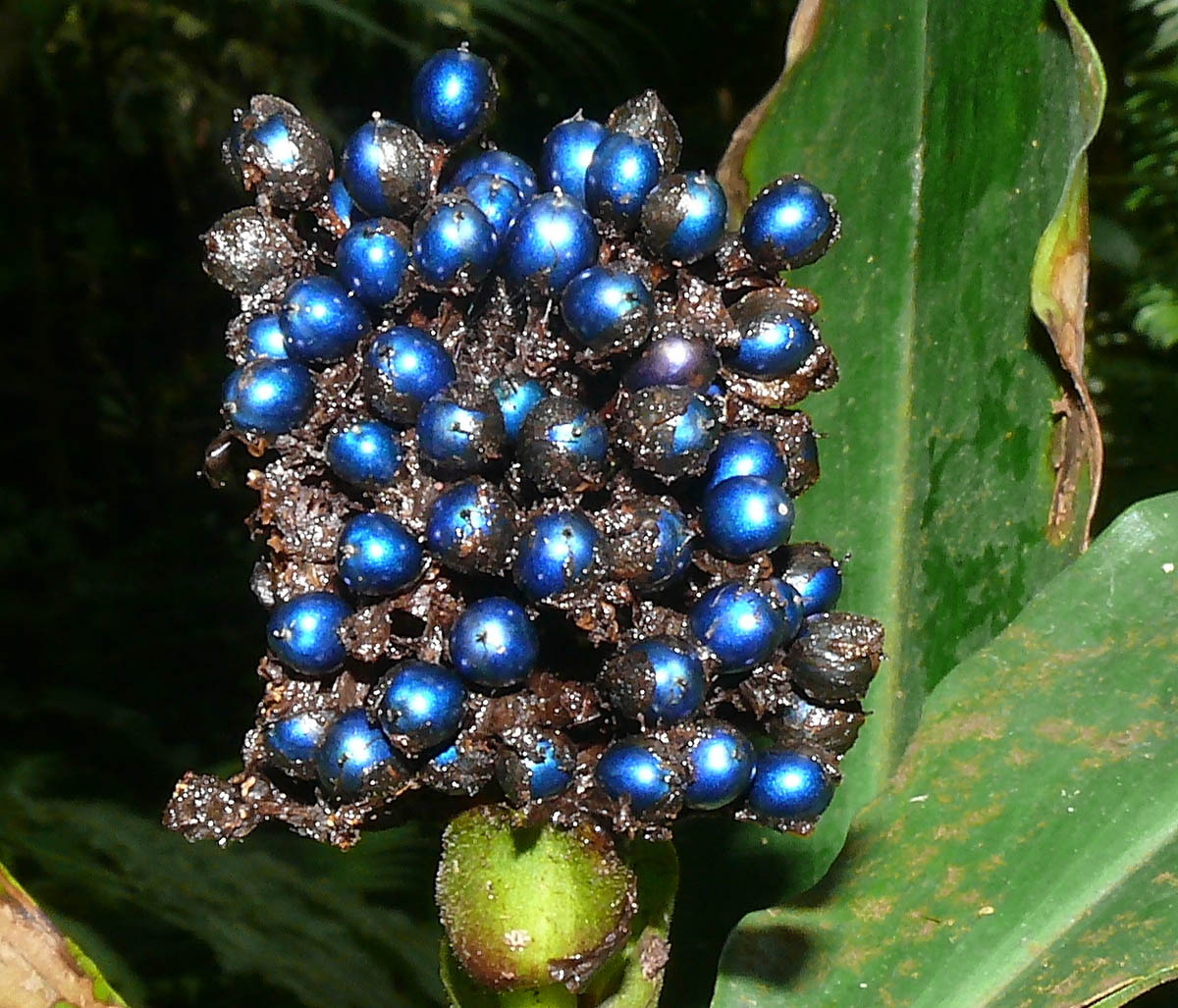
Pollia condensata fruit, photographed 8 July 2010. A contextual example, not the preserved Ghanaian specimen measured in the 2012 study. Juliano Costa · CC BY-SA 3.0

Research & assessmentOriginal paper · PNAS issue 25 September 2012; online reporting 11 September 2012

The blue fruit that keeps its colour without blue pigment

Twisted layers of cellulose give Pollia condensata fruit a metallic, pointillist colour that can persist in old specimens.

The fruit of Pollia condensata can look like a cluster of tiny metallic beads. A 2012 investigation led by Silvia Vignolini traced its brilliant blue appearance to structures in the cell walls, rather than an extracted blue pigment.

Layers of cellulose microfibrils twist within the outer cells. Their arrangement selectively reflects light. Differences between neighbouring cells produce different reflected colours, so the fruit has a speckled, almost pointillist appearance rather than one uniform painted surface.

The paper includes fruit collected in Ghana in 1974 and preserved at Kew. Its continued colour illustrates a consequence of the mechanism: the appearance can survive after the fruit dries, because the reflecting structure remains.

The authors also discuss possible roles in seed dispersal, but those proposals are separate from their optical measurements. The paper is offered for its anatomical and physical explanation. It does not establish that every brightly coloured fruit uses the same mechanism, or that an old claim of exceptional brightness is a permanent world record.

Source details & what was checked
  • Silvia Vignolini, Paula J. Rudall, Alice V. Rowland, Alison Reed, Edwige Moyroud, Robert B. Faden, Jeremy J. Baumberg, Beverley J. Glover and Ullrich Steiner · PNAS issue 25 September 2012; online reporting 11 September 2012
    Cambridge-hosted four-page author paper PDF inspected: abstract, anatomical/optical explanation, Fig1 provenance and hypothesis wording. Supporting information not reviewed; bright fruit is not a claim of current world-record status. Writer meaningful web reading; desktop/mobile reader QA remains pending.
    Pointillist structural color in Pollia fruit (external source, opens a new tab) · In the source library
A small brown lizard with a curling tail and pale side markings rests on a broad green leaf.
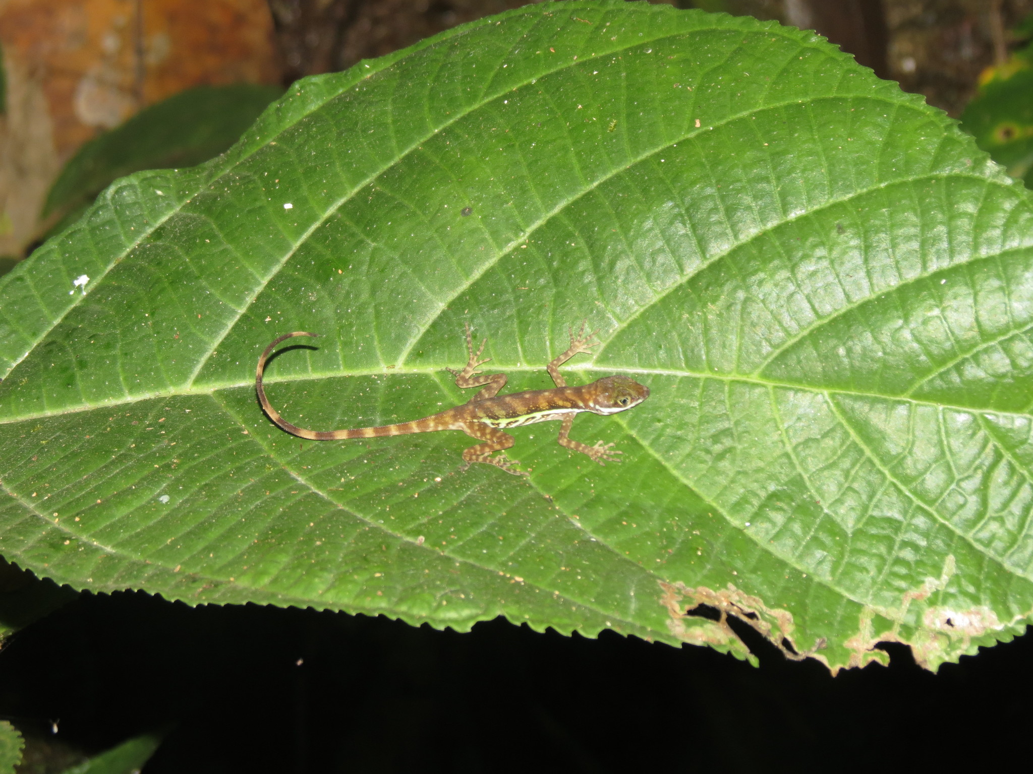
Reuven Martin photographed this water anole on a leaf in Costa Rica on 4 February 2018. The photograph shows the species above water, separate from the diving trials. Reuven Martin / iNaturalist via Wikimedia Commons · CC0 1.0 Universal

Research & assessmentOriginal paper · 18 September 2024 (Biology Letters and PMC citation)

The lizard that breathes from its own bubble

A water anole carries exhaled air on its head. A field experiment measured how much longer it could stay underwater with the bubble intact.

Added · Daily discovery

Watch the snout of a submerged water anole. A bubble swells against the lizard’s skin, then shrinks until it disappears. The lizard has drawn the air back in. In photographs from Lindsey Swierk’s 2024 study, the bubble sits either on top of the snout or beside it, between the eye and nostril.

Water anoles live along rocky streams in parts of Costa Rica and Panama. They can dive to escape predators. Tiny structures on the skin of semi-aquatic anoles help hold a film of air against the body. Swierk wanted to know what difference the bubbles made to the time these lizards could spend out of sight.

She compared two randomly assigned groups in a streamside tank in Costa Rica. An emollient interrupted normal bubble formation in one group, with the nostrils left clear. The other group received water. Each trial ended when the animal surfaced. Of the 28 completed trials, one unusually long control dive was excluded from the analysis. Swierk reported that lizards with normal bubbles stayed down an average of 67.5 seconds longer.

A follow-up asked where the bubble’s oxygen comes from. Alexandra M. Martin, Diane Cordero-De La Cruz and Swierk varied the amount of oxygen dissolved in the tank water. The lizards dived longest in the highest-oxygen treatment. That pattern is consistent with oxygen passing from the water into the bubble. In her account of the study, Swierk identifies directly measuring that transfer as a next step.

Start with Figure 1 of the 2024 paper. The first four frames show the same bubble at intervals of a fifth of a second. The sequence puts a visible action behind the extra time measured by the stopwatch.

The second source is a freely readable explanation by a study coauthor. The follow-up measured dive duration in water with different dissolved oxygen levels.

Source details & what was checked
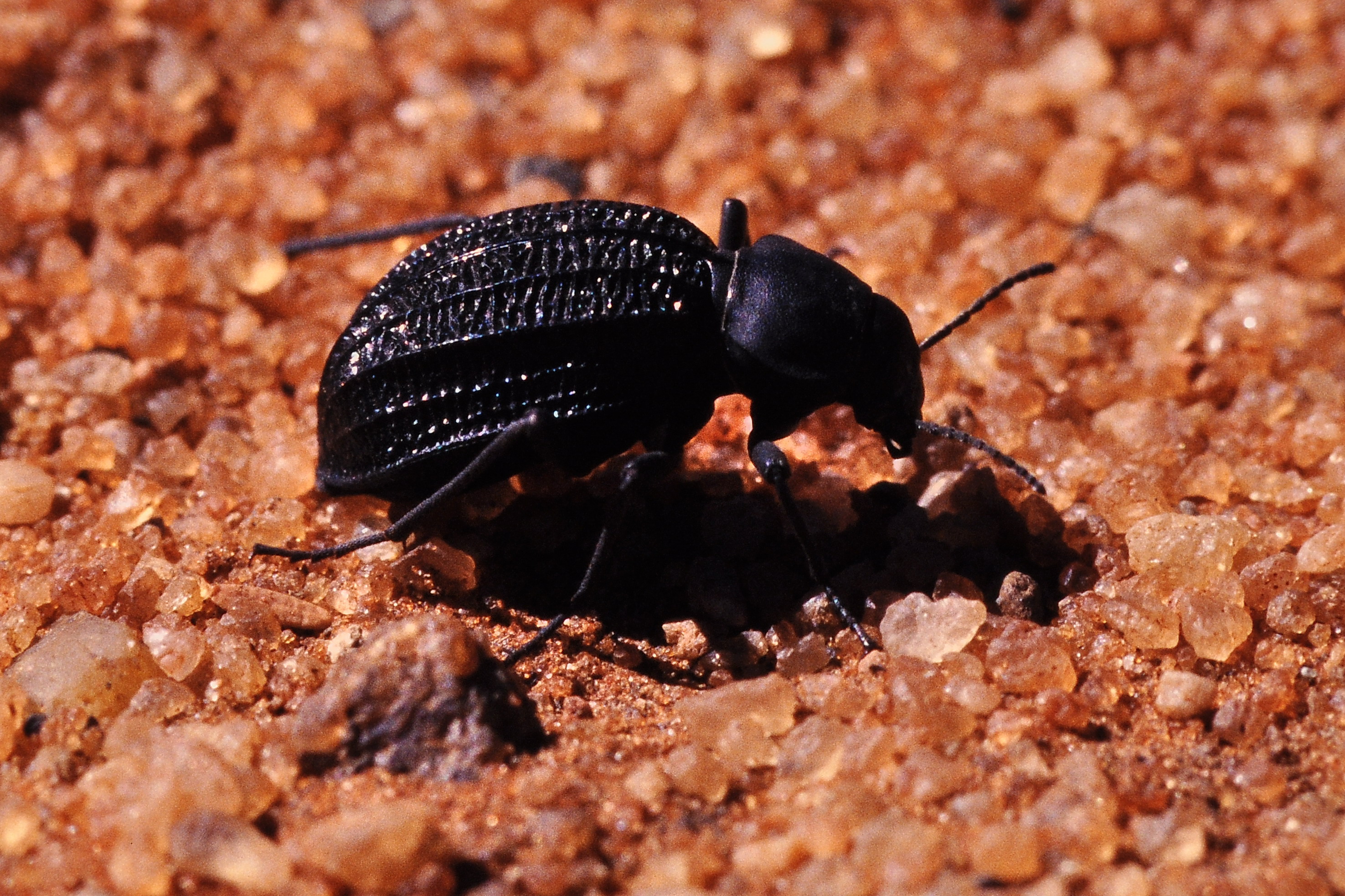
A glossy black beetle with lengthwise grooves across its back stands on coarse orange sand.
A Namib fog-basking beetle on sand, photographed by Schnobby on 22 March 2012. The separate laboratory photograph in the lead paper shows the head-down drinking stance. Onymacris unguicularis in Namib Desert by Schnobby, via Wikimedia Commons, CC BY-SA 3.0 · Creative Commons Attribution-ShareAlike 3.0 Unported

Research & assessmentOriginal paper · 16 July 2010. Frontiers in Zoology 7, article 23.

The beetle that tips forward to drink fog

On Namib dunes, a darkling beetle lowers its head and lets droplets that land on its back run towards its mouth.

Added · Daily discovery

A fog-basking beetle can drink without finding a puddle. The Namib darkling beetle Onymacris unguicularis climbs to a dune ridge and lowers its head. Tiny droplets gather on its wing cases and trickle down towards its mouth.

In a 2010 study, Thomas Nørgaard and Marie Dacke brought the behaviour into a fog chamber. A live beetle in Figure 4 is leaning much less steeply than the term “headstand” suggests. The underside of the body averaged about 23 degrees from horizontal. The other three beetle species kept moving throughout their 20-minute observations.

A separate study had proposed a water-catching mechanism based on a bumpy shell. In 2003, field researchers William J. Hamilton III, John R. Henschel and Mary K. Seely argued that its photograph showed a different beetle from the one named. In the 2010 comparison, the head-down drinker had relatively smooth, grooved wing cases. The authors found the stance and the climb to the ridge more important to its use of fog than the precise texture of its back.

A follow-up chamber experiment compared dead beetles mounted in the drinking position with lengths of dune bushman grass. The grass yielded more water over two hours. Drops ran off the beetle specimens more steadily. The researchers suggested that a steady supply could help a live beetle get a drink even during a short spell on an exposed ridge.

Source details & what was checked
  • Thomas Nørgaard and Marie Dacke · 16 July 2010. Frontiers in Zoology 7, article 23.
    Read introduction, behavioural methods and results, passive fog collection results, discussion and conclusions. Viewed the actual authors’ Figure 4 PNG, which shows the live beetle and annotated posture angle. The clean anonymous browser URL displayed the relevant article body at all three widths. Fifteen tested O. unguicularis performed the stance in this cold fog chamber. Three comparison species did not. 22.7° is the mean ventral body angle to horizontal. These tests do not establish that surface structure never matters, or that O. unguicularis is the only species in the desert that fog-basks.
    Fog-basking behaviour and water collection efficiency in Namib Desert Darkling beetles (external source, opens a new tab) · In the source library
  • William J. Hamilton III, John R. Henschel and Mary K. Seely · March/April 2003. South African Journal of Science 99, p. 181.
    Read the single PDF page, including the complete beetle letter in the lower half, its journal header and author lines. The upper part finishes an unrelated geology reference list. The archive filename contains “Namid”; the printed title correctly says Namib. The authors argued that the photo appeared to depict Physosterna cribipes, a different beetle. They did not dispute the possibility of textured artificial or beetle surfaces collecting fog, and the draft does not resolve the identity of every specimen used by the earlier authors.
    Fog collection by Namib Desert beetles (external source, opens a new tab) · In the source library
  • Thomas Nørgaard, Martin Ebner and Marie Dacke · 3 April 2012. PLOS One 7(4), e34603.
    Read the full open article’s materials, results, Figure 3 and Figure 4 descriptions, and discussion. The separate duration trial sampled at 20-minute intervals. Normal Chrome opened the exact article route at all three tested widths. The comparison was between dead beetles mounted head down, grass segments and, for the final amount experiment, wire controls. Beetle runoff was comparatively steady in the chamber. Whether living beetles derive the same benefit in the wild is an inference made by the authors, not a result this comparison established.
    Animal or Plant: Which Is the Better Fog Water Collector? (external source, opens a new tab) · In the source library
Back to sections ↑

Borrowed parts, living animals

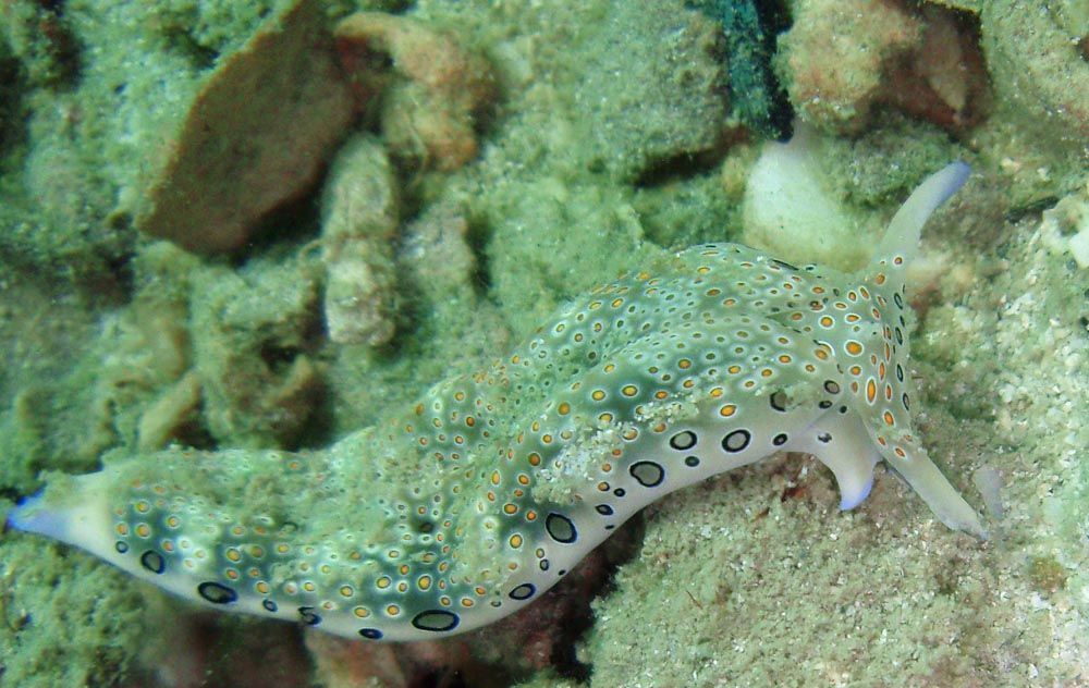
A sea slug photographed underwater at Koh Phangan.
A sea slug identified by its Commons record as Plakobranchus ocellatus, photographed at Koh Phangan, Thailand, in January 2014 by Chaloklum Diving. A contextual field photograph; neither the experimental PoB lineage nor the photographed individual’s genotype is established by this image record. Chaloklum Diving · CC BY 3.0 Unported

Research & assessmentOriginal paper ·

The sea slug that keeps working parts of its food

A sea slug retains algal chloroplasts; a genome study tested how this borrowed photosynthesis survives without borrowed nuclear genes.

Edition · Daily discovery

Backfilled edition · First published .

A sea slug can digest algae while keeping some of their working machinery. In a process called kleptoplasty, chloroplasts taken from food remain inside the animal’s intestinal cells. These are the structures that carry out photosynthesis. In some sea slugs they continue working for months, long after the rest of the meal has disappeared.

Taro Maeda and colleagues investigated Plakobranchus ocellatus in a 2021 eLife paper. They studied the lineage they call type black, or PoB: the familiar species name covers a group of closely related animals. Their measurements included oxygen production, photosynthetic efficiency and survival under different lighting conditions. The retained chloroplasts remained active after more than three months without feeding. Slugs given alternating light and darkness lived longer, on average, than those kept continuously dark.

The genetic result addressed a different puzzle. Chloroplasts normally depend on proteins whose genes sit in the algal nucleus. A proposed explanation was that slugs had acquired the necessary algal genes. As Science Japan explains, the team searched the slug genome, raw sequencing data and gene-expression data without finding evidence for the photosynthesis-related transfers they were looking for. That result did not mean the entire maintenance mechanism had been solved.

Keio University’s project page places the work within research on traits acquired through retained organelles and proteins. For this animal, the useful distinction is between keeping a chloroplast and inheriting an alga’s nuclear instructions. The open paper supplies the experimental details; the Science Japan account explains why obtaining good-quality slug DNA mattered to the investigation.

Source limitations and contextual photograph distinctions remain as recorded.

Source details & what was checked
  • Taro Maeda, Shunichi Takahashi, Takao Yoshida, Shigeru Shimamura, Yoshihiro Takaki, Yukiko Nagai, Atsushi Toyoda, Yutaka Suzuki, Asuka Arimoto, Hisaki Ishii, Nori Satoh, Tomoaki Nishiyama, Mitsuyasu Hasebe, Tadashi Maruyama, Jun Minagawa, Junichi Obokata and Shuji Shigenobu ·
    Meaningful selective full-text reading: abstract, introduction, Figure1 context, Results on light/oxygen/survival, genome-search discussion and Figure5 candidate gene discussion. Supplementary raw data and all methods not independently reanalysed. Free article body and CC BY notice visible; optional PMC account controls are not required for reading. Reading route independently rechecked on 6 November 2026 at desktop, 390px and 320px; the original content-reading and interpretation limits remain.
    Chloroplast acquisition without the gene transfer in kleptoplastic sea slugs, Plakobranchus ocellatus (external source, opens a new tab) · In the source library
  • Science Japan / Japan Science and Technology Agency; translated with permission from The Science News Ltd.; no individual byline shown ·
    Complete main text read, including genotype-search account and explicit statement that the molecular maintenance mechanism remained unknown. This is an institutional news explanation, not a second experiment. Photographs on this page explicitly prohibit unauthorised reproduction and are not selected for reuse. Reading route independently rechecked on 6 November 2026 at desktop, 390px and 320px; the original content-reading and interpretation limits remain.
    Genome sequencing of Plakobranchus ocellatus: An animal that conducts photosynthesis (external source, opens a new tab) · In the source library
  • Institute for Advanced Biosciences, Keio University; Taro Maeda project listing · Undated; inspected 4 November 2026
    Complete short project description and publication list read, including link to the 2021 eLife paper. Project framing, not independent confirmation of the experiment. Reading route independently rechecked on 6 November 2026 at desktop, 390px and 320px; the original content-reading and interpretation limits remain.
    Kleptobiolgy Project (external source, opens a new tab) · In the source library
Back to sections ↑

Homes aligned with the sun

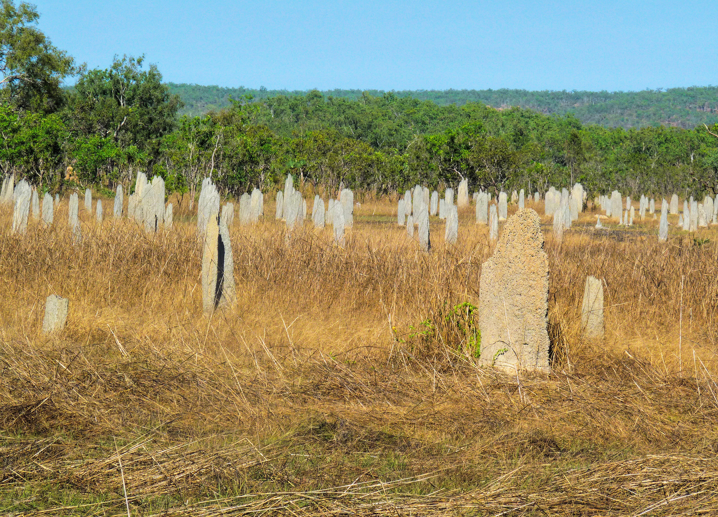
Flat pale termite mounds standing in dry grass beneath a blue sky in Litchfield National Park
Magnetic termite mounds in Litchfield National Park, Northern Territory, photographed 5 July 2009. A later contextual view of the aligned structures, separate from the Archives’ 1950 photograph. Ianperegian, via Wikimedia Commons · CC BY-SA 4.0 International

History & recordFirst-party history and archival caption · 2010 learning-resource text; photograph 1950

The termite homes that line up north to south

Northern Australia’s magnetic termite mounds present broad faces to east and west, with temperature shaping their orientation.

Edition · Daily discovery

Backfilled edition · First published .

A field of termite mounds in northern Australia can seem to share a common alignment. The thin edges run roughly north to south, while the broad faces look east and west. These are the homes of the grass-eating termite Amitermes meridionalis, often called magnetic or compass termites.

The National Archives of Australia introduces the structures through a 1950 photograph. A person stands among nine towers, making their scale easier to read. Its explanation connects the alignment with morning and evening warming and reduced midday exposure. Wind can alter the direction; the field pattern need not be perfectly uniform.

Seasonal flooding gives height a separate importance. The Archives notes that low sections can be inundated for weeks, leaving the above-ground home especially important. Its account reports temperature differences of up to eight degrees Celsius between sunlit and shaded surfaces.

The Northern Territory’s two-page Nature Notes places these structures among very different termite homes. Tall cathedral mounds belong to Nasutitermes triodiae; other species build in trees or remain underground. The guide describes termites breaking down dead plant material with help from microorganisms in their guts. Their mounds store nutrients which return to the soil as an abandoned mound breaks down. Their excavations in trees also provide habitat for other animals, including birds, bats and reptiles.

The word magnetic needs care. The Archives describes use of Earth’s magnetic field as a speculation in its account. That possible directional cue is a separate question from the temperature advantage of an alignment. Seeing the structures does not establish how the insects choose their direction.

The 2010 archival learning resource calls magnetic-field guidance a speculation; this article does not turn that historical account into a current consensus claim. The photograph is a separate 2009 contextual view, not the archival 1950 image.

Source details & what was checked
  • Education Services Australia Limited and National Archives of Australia · 2010 learning-resource text; photograph 1950
    Complete archival learning-resource main body and educational-value notes independently read in the actual browser on 5 November 2026 at desktop, 390px and 320px. The learning text is dated 2010 and interprets a 1950 photograph. It explicitly labels geomagnetic guidance as speculation; no primary orientation or magnetoreception experiment was independently read, and this historical account is not presented as a 2026 consensus review. Minor narrow-screen overflow requires ordinary panning on the source site. Reading route independently rechecked on 6 November 2026 at desktop, 390px and 320px; the original content-reading and interpretation limits remain.
    Magnetic termite structures in the Northern Territory (external source, opens a new tab) · In the source library
  • Stuart Traynor and Dean McAdam; illustrations Bob Whiteford; Northern Territory Government · Undated two-page government nature guide
    Both pages independently read in the ordinary native PDF reader on 5 November 2026, including authors, mound-type distinctions, nutrition and tree-hollow habitats. The published legacy NT URL redirects normally to the same freely readable guide at https://nt.gov.au/_media/docs/parks/parks-resources-for-schools/nature-notes/invertibrates/termite-homes.pdf. Desktop, 390px and 320px native reading/panning exercised without login, payment or a required app. Further-reading book mentions are not recommended purchases. Browser widths were tested, not physical devices. Reading route independently rechecked on 6 November 2026 at desktop, 390px and 320px; the original content-reading and interpretation limits remain.
    Nature Notes — Termite homes / Termites: Nature’s recycling agents (external source, opens a new tab) · In the source library
Back to sections ↑